Home

przerwać konserwatywny dźwignia cd86 m1 marker Amazon mistrz Letni I

M1-EVs treatment for 24 h induced expression of iNOS and CD86 in RAW... |  Download Scientific Diagram
M1-EVs treatment for 24 h induced expression of iNOS and CD86 in RAW... | Download Scientific Diagram
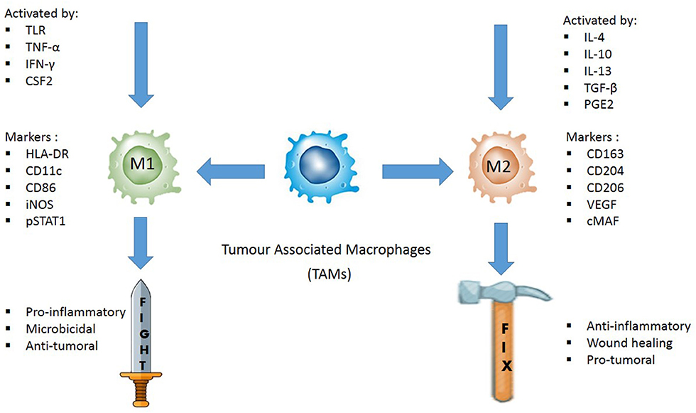
Frontiers | Evaluating the Polarization of Tumor-Associated Macrophages  Into M1 and M2 Phenotypes in Human Cancer Tissue: Technicalities and  Challenges in Routine Clinical Practice
Frontiers | Evaluating the Polarization of Tumor-Associated Macrophages Into M1 and M2 Phenotypes in Human Cancer Tissue: Technicalities and Challenges in Routine Clinical Practice
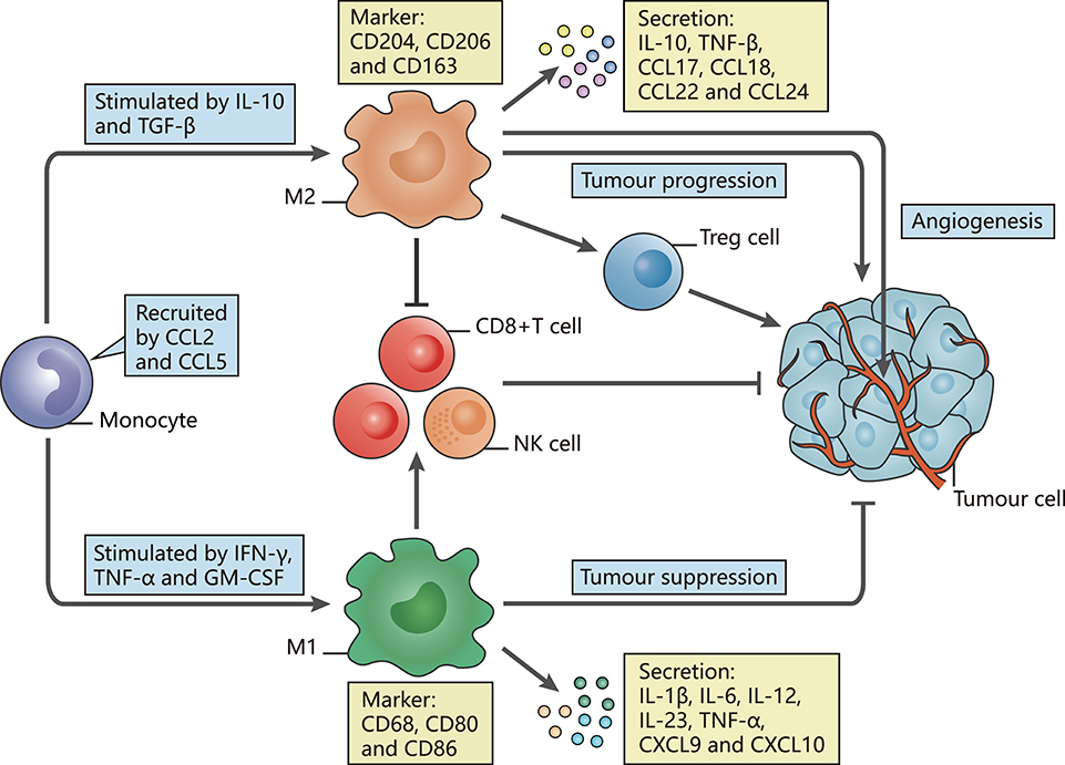
Frontiers | Redefining Tumor-Associated Macrophage Subpopulations and  Functions in the Tumor Microenvironment
Frontiers | Redefining Tumor-Associated Macrophage Subpopulations and Functions in the Tumor Microenvironment

Immunofluorescence staining of M0, M1, and M2 macrophage cultures after...  | Download Scientific Diagram
Immunofluorescence staining of M0, M1, and M2 macrophage cultures after... | Download Scientific Diagram

HCV core protein inhibits polarization and activity of both M1 and M2  macrophages through the TLR2 signaling pathway | Scientific Reports
HCV core protein inhibits polarization and activity of both M1 and M2 macrophages through the TLR2 signaling pathway | Scientific Reports

M1 and M2 Macrophage Markers | OriGene

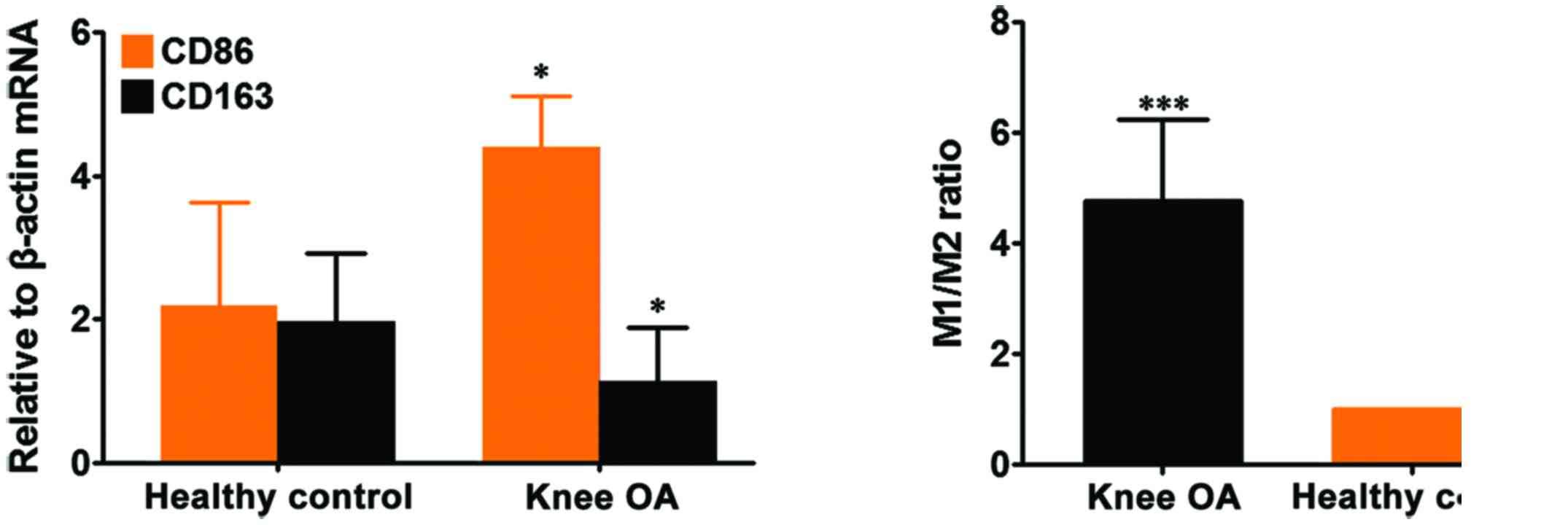
Imbalance of M1/M2 macrophages is linked to severity level of knee  osteoarthritis
Imbalance of M1/M2 macrophages is linked to severity level of knee osteoarthritis

M1 and M2 Macrophage Markers | OriGene

In vitro M1 to M2 polarization of macrophages induced by PDA/MTX@TSG.... |  Download Scientific Diagram
In vitro M1 to M2 polarization of macrophages induced by PDA/MTX@TSG.... | Download Scientific Diagram

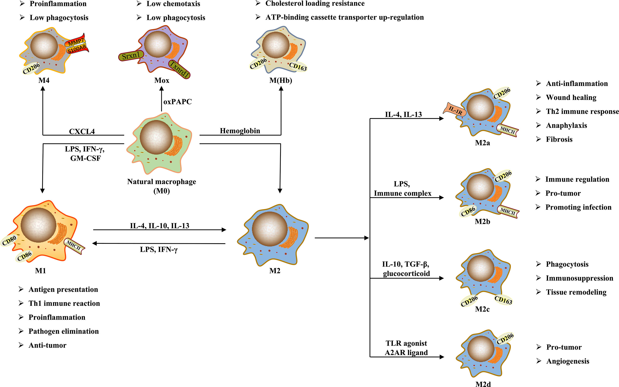
Frontiers | Macrophage Polarization and Its Role in Liver Disease

Flow cytometry analysis of M1 and M2 surface markers (A) The... | Download  Scientific Diagram
Flow cytometry analysis of M1 and M2 surface markers (A) The... | Download Scientific Diagram

CD86, MHCII, and CD206 identify M1-and M2-like cells in in vitro... |  Download Scientific Diagram
CD86, MHCII, and CD206 identify M1-and M2-like cells in in vitro... | Download Scientific Diagram

Transcription factor KLF4 regulated STAT1 to promote M1 polarization of  macrophages in rheumatoid arthritis | Aging
Transcription factor KLF4 regulated STAT1 to promote M1 polarization of macrophages in rheumatoid arthritis | Aging

B7-2/CD86 Antibody (BU63) (NBP2-25208): Novus Biologicals

Polarization of M1 and M2 Human Monocyte-Derived Cells and Analysis with  Flow Cytometry upon Mycobacterium tuberculosis Infection
Polarization of M1 and M2 Human Monocyte-Derived Cells and Analysis with Flow Cytometry upon Mycobacterium tuberculosis Infection

M2 but not M1 macrophages promote cancer metastasis. A, FACS analysis... |  Download Scientific Diagram
M2 but not M1 macrophages promote cancer metastasis. A, FACS analysis... | Download Scientific Diagram
M1 Macrophage Flow Cytometry Panel: R&D Systems

a) Flow cytometric analysis of CD206 + M2-macrophages and CD86 +... |  Download Scientific Diagram
a) Flow cytometric analysis of CD206 + M2-macrophages and CD86 +... | Download Scientific Diagram

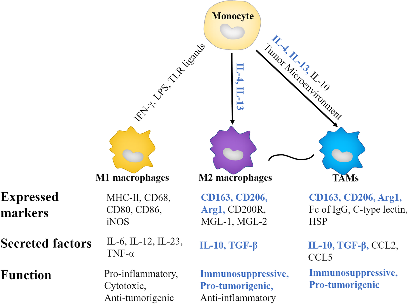
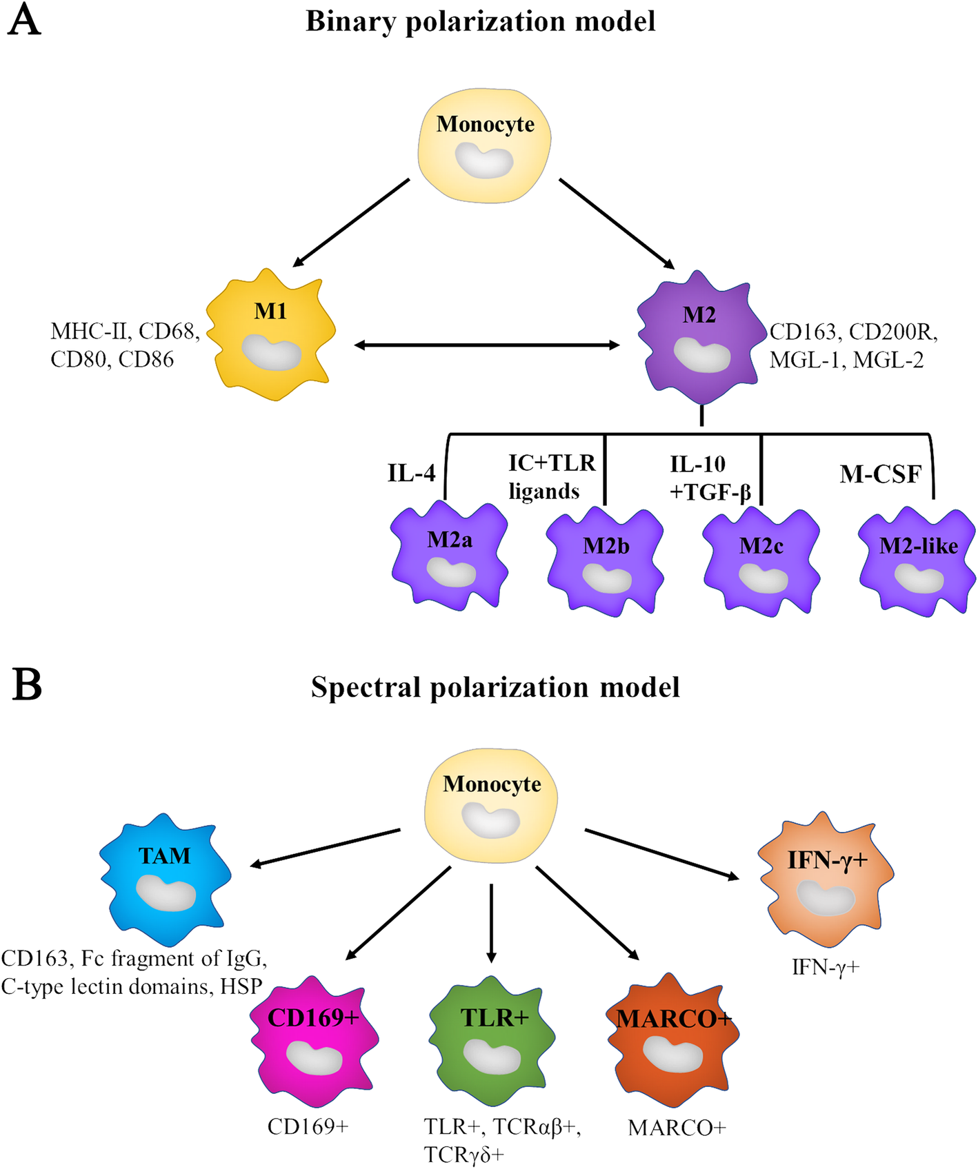
Tumor-associated macrophages: an accomplice in solid tumor progression |  Journal of Biomedical Science | Full Text
Tumor-associated macrophages: an accomplice in solid tumor progression | Journal of Biomedical Science | Full Text
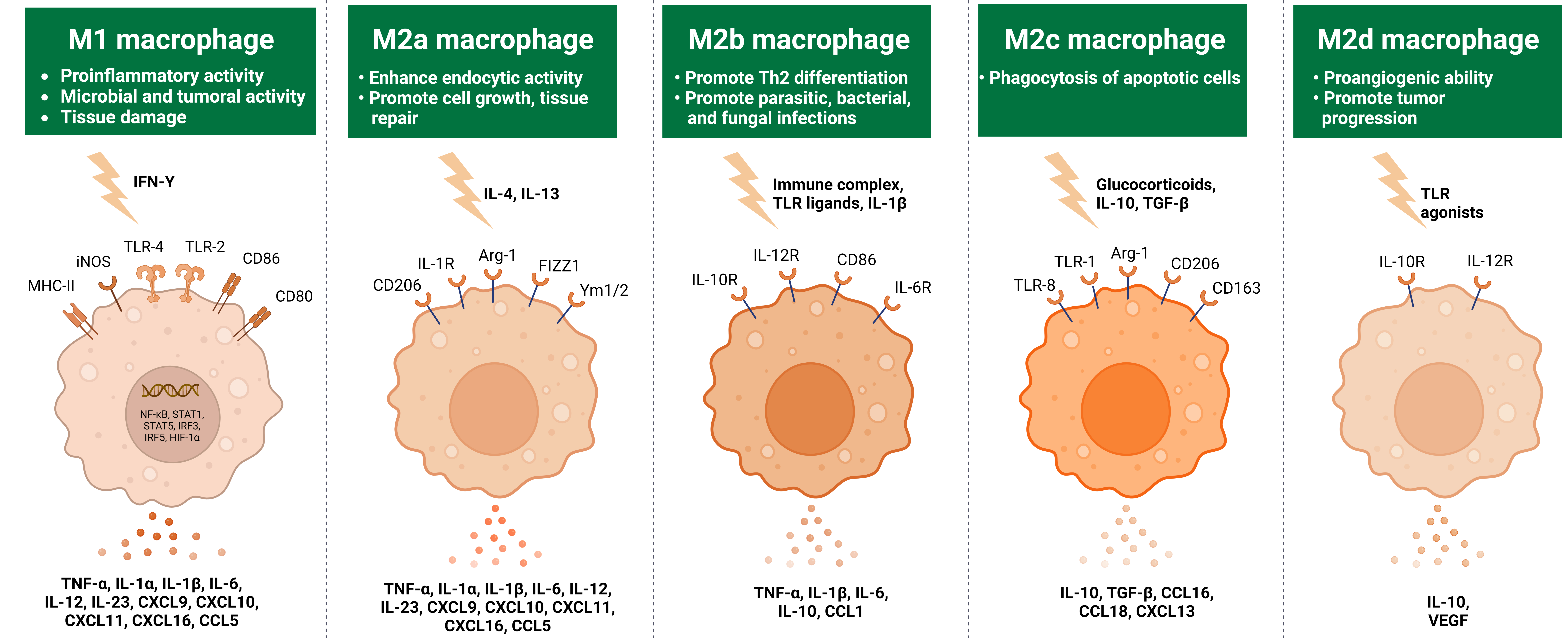
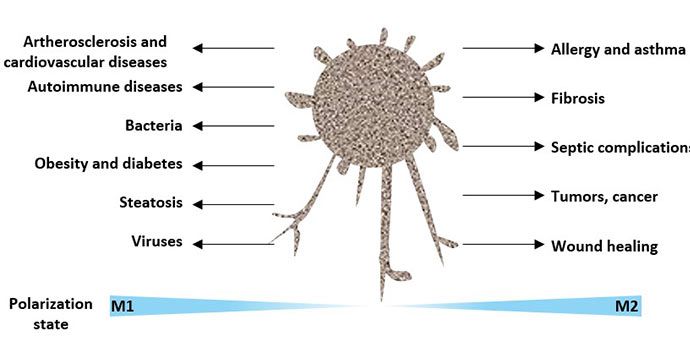
Two Types of Macrophages: M1 and M2 Macrophages - Cusabio

xmlinkhub
xmlinkhub

Tumor-associated macrophages: an accomplice in solid tumor progression |  Journal of Biomedical Science | Full Text
Tumor-associated macrophages: an accomplice in solid tumor progression | Journal of Biomedical Science | Full Text
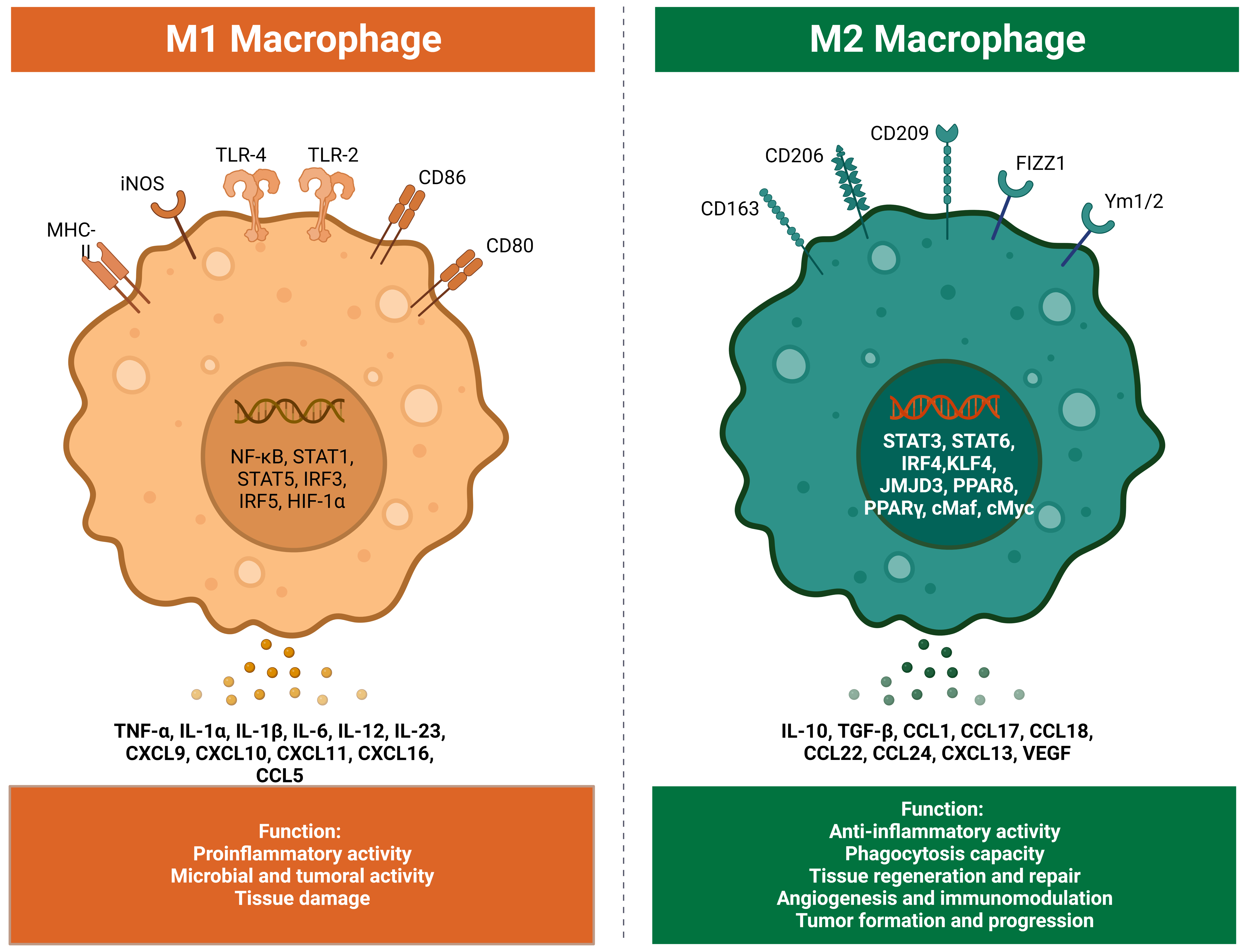
CD86 as the marker of M1 was detected by flow cytometry. (A) Flow... |  Download Scientific Diagram
CD86 as the marker of M1 was detected by flow cytometry. (A) Flow... | Download Scientific Diagram